Present position:
 LOIS-3D
NIR-I & NIR-II In vivo 3D Optoacoustic Imaging System
The LOIS-3D system can achieve NIR-I & NIR-II optoacoustic imaging effects because its excitation wavelength can reach 660-2300 nm. It can perform 2D and 3D imaging of the biological characteristics of living animals and obtain qualitative and quantitative data, providing more accurate diagnostic analysis for drug targeted therapy, early tumor screening, cardiovascular disease and brain disease monitoring and other research.
- Composition
- Application
- Advantage
- Cooperation
-
LOIS-3D system laser
 Using the well-known EKSPA PhotoSonus series, with high energy excitation, high throughput wavelength output, fast wavelength coordination, mobility and other advantages, to provide high quality imaging data for in vivo or tissue studies
- High energy output set pumped laser with 180 MJ standard and 250 MJ higher, integrated OPO and PSU
- Wavelength tuning ranges from 660-1064 nm, and 660-2300 nm for higher configurations, covering NIR-I & NIR-II
- 4 laser beams (2 orthogonal, 2 oblique) are simultaneously excited to ensure the uniform energy received by the tissue
LOIS-3D system imaging mode
The model of LOIS-3D / LOIS-3D Plus produced by TomoWave Laboratories is the whole body 3D optoacoustic imaging system specially designed for living animals in the NIR-I & NIR-II
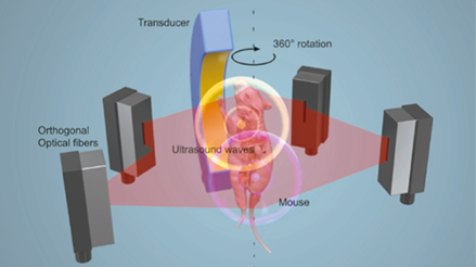
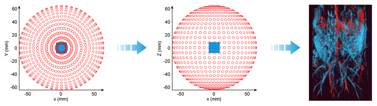
- Small animal is fixed on a special holder, driven by a precision rotating motor to complete a 360° rotation
- 4 short pulse lasers (two orthogonal and two oblique) are emitted by laser lasers
- The tissue absorbs light energy to generate an ultrasonic signal, which is received by a professionally designed 120° arc transducer
- Collect signals at 360° and adopt unit conversion through DAQ data, dedicated 3D reconstruction software reconstructs optoacoustic images
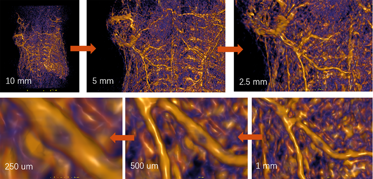
LOIS-3D system technology advantages
- Imaging wavelength 660-1064 nm / 660-2300 nm, NIR-I & NIR-II full-band continuous imaging
- X-Y-Z isotropic resolution of 3D space is 150 μm
- Light absorption contrast 0.03 [1/cm] (~1pmole of ICG)
- Full field imaging of mouse body and brain (40 mm x 40 mm x 40 mm)
- Imaging depth > 4.5cm@mouse
-
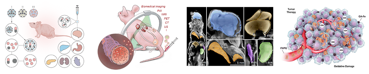
Can realize 3D optoacoustic imaging of small animals in the NIR-I & NIR-II (660-2300 nm), high-resolution imaging of blood vessel morphology, and highly specific functional detection of different tissue components. Realizes multi-scale tracing and functional imaging from cells to tissue, and has important applications in many biomedical fields, such as molecular probes, bio-nanomaterials, cardiovascular diseases (angiogenesis, myocarditis, thrombosis, myocardial infarction,etc.), hemoglobin monitoring, early tumor detection and efficacy monitoring, sentinel lymph node monitoring, brain imaging and brain function monitoring and other frontier research.
-
Development of bio-nano materialsDevelopment and application ofphotothermographic agentsDevelopment and applicationof bio-nano materialsBiodistribution of nanoprobesBlood concentration…
-
Targeted tumor imagingSubcutaneous tumorIn situ tumors of the brainOrthotopic tumor of lower extremityProstate tumor…
-
Anatomy imagingOrgans and spineCardiovascular SystemBlood vessel microstructureBrain structure and blood vessels…
-
Synergistic treatmentOptoacoustic cavitationPhotothermal and AcousticDynamicsOptoacoustic and photochemistry…
-
-
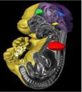
Unlabeled optoacoustic imaging --Cerebral vascular imaging
Brain hemoglobin imaging can be used to detect brain tumors and traumatic brain injury (traumatic brain injury), and to monitor and evaluate the efficacy of cerebrovascular leakage, such as cerebral amyloidosis, stroke, etc.
 OptoAcoustic Imaging of Live Mouse Brain,Total Hemoglobin MapR. Su et al. J. Biomed. Opt. 2012Brain @1064nmLower Brain and Neck @760 nm
Unlabeled optoacoustic imaging --Microscopic vascular structure imaging
Microscopic hemoglobin angiography allows the observation of hematoma lesions and possible lesions and plaques in the surrounding area to detect cardiovascular diseases, such as atherosclerosis, myocarditis, thrombosis, myocardial infarction, etc.
Labeled optoacoustic imaging--bio-nanoprobe tumor imaging
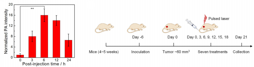
The use of organic NIR-II photosensitizer targeting tumor probe 1064nm for tumor imaging can assist the diagnosis and treatment of preclinical tumor diseases.
Tumor imaging at different time pointsSubcutaneous tumor Post-injection 12hMouse body with high resolution detailsBrain vasculature can be visualized
in a live mouse with scull and skin intact
-
TomoWave has been highly recognized by users since the introduction of the LOIS-3D NIR-I & NIR-II In vivo 3D Optoacoustic Imaging System. So far, it has been installed in the MD Anderson Cancer Research Center, Washington University in St. Louis, University of Houston, Qingdao University, Guangxi University and so on. We have cooperated with many scientific research teams such as Shenzhen Institute of Advanced Technology, Chinese Academy of Sciences, Sun Yat-sen University, the Third Affiliated Hospital of Sun Yat-sen University, Huazhong University of Science and Technology, Soochow University, South China Normal University, Huazhong Agricultural University, Nanjing University of Technology, Nanjing University of Posts and Telecommunications, etc. The in vivo optoacoustic imaging characterization of the study was tested on the LOIS-3D NIR-I & NIR-II whole-body 3D optoacoustic imaging system for small animals.
NIR-I,non-photosensitive polymer nanovesicles for tumor therapy
Angew.Chem.Int.Ed. reported on a non-photosensitive material under the irradiation of pulsed laser to produce highly reactive substances, for cancer diagnosis and treatment. The fluorescence/optoacoustic dual-mode imaging of TomoWave's 3D optical system provides precise guidance for in-vivo pulsed laser therapy.


 Angew. Chem. Int. Ed. 60,(2021),4720-4731Non-photosensitive polymer nanosicles (NO-NCPs) have the effect of optoacoustic cavitation to enhance ROS, and can accelerate the release of NO in acid lysosomes, thus providing highly reactive peroxide nitrite ONOO-, which promotes mitochondrial damage and DNA fragmentation of cancer cells, and thus apoptosis.
NIR-II,Study on tumor targeting of optoacoustic contrast agents
Small published a synthesis method of mini hollow gold nanorods (M-AuHNRs) with strong plasma absorbance. Under the same optical density, excited by TomoWave optoacoustic imaging system NIR-II, the optoacoustic signal intensity is 3.5 times stronger than that of ordinary hollow gold nanorods.


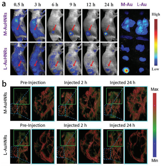 Miniature hollow gold nanorods (M-AuHNRs) (length≈46 nm) can enhance the effect of optoacoustic imaging in the NIR-II window Small 16, (2020), 37
NIR-II, Photothermal and Acoustic Dynamic Synergy Therapy
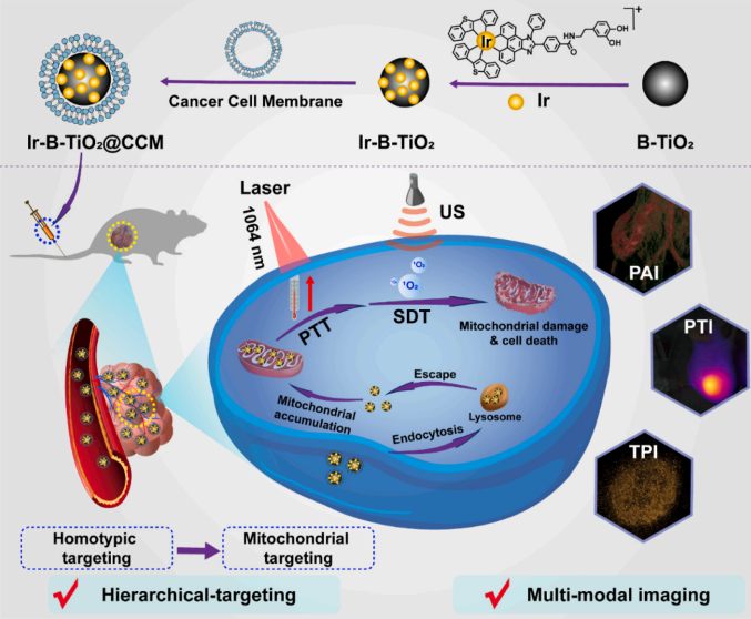
Biomaterials reported a collaborative treatment method in NIR-II photothermal irradiation and ultrasound. Ir-B-TiO2@CCM realizes the tracking of the circulation of nanoparticles in the body under the mediation of multi-modal imaging, thereby monitoring the temperature change of the tumor during the treatment, and finally effectively inhibiting the tumor in the mouse animal model experiment metastasis and recurrence.


 The characteristics of homologous targeting and immune escape of cancer cell membrane promote the selective accumulation of Ir-B-TiO2@CCM to homologous tumor cells and avoid immune rejection of macrophages at the same time, so as to improve tumor accumulation and treatment safety; The results in vivo and in vitro showed that the synergistic treatment of PTT and SDT had more superior tumor inhibition.Biomaterials 275, (2021), 120979