COMPANY HISTORY

TomoWave Laboratories, Inc. was founded in 2010 by Dr. Alexander A. Oraevsky, the father of biomedical optoacoustic imaging in the world. It is the pioneer of biomedical optoacoustic imaging technology in the world. Focusing on oncology and biology, our laboratory is committed to the development and transformation of high-resolution, high-contrast, and high-sensitivity 3D optoacoustic tomography (OAT),Diagnosis and treatment and hemoglobin applications mediated by new functional nanomaterials as an integrated diagnosis and treatment platform play a leading role. Over 10 years of experience in pioneering research in the field of optoacoustic imaging, sensing and monitoring.
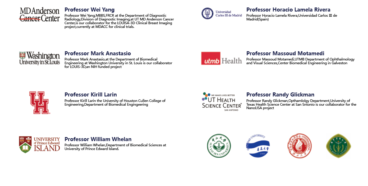
TomoWave Suzhou was founded in November 2020, with the participation of Jiangsu Industrial Technology Research Institute and Taicang Biomedical Industrial Park and Taicang Innovation Investment and Development Co., LTD. It is an important part of the province's scientific research innovation system. It is committed to transforming the preclinical animal optoacoustic imaging system and clinical breast cancer optoacoustic diagnosis system in China. TomoWave system has been installed at the MD Anderson Cancer Center, Washington University in St. Louis, the University of Texas At SAN Antonio Health Science Center, the University of Houston, Guangxi University, Qingdao University, etc.
THE CORE TECHNOLOGY
Optoacoustic imaging is an emerging noninvasive hybrid imaging technology that combines optical excitation and ultrasonic detection. During the imaging process, the optoacoustic contrast agent absorbs a non-ionizing pulse laser and converts it into heat, which generates sound waves through thermoelastic expansion. These sound waves are detected using broadband ultrasonic transducers and optoacoustic images can be constructed according to the arrival time of the sound waves. Compared with photons, phonons scatter less in biological tissues, so optoacoustic imaging has the advantages of high tissue contrast, high spatial resolution and deep imaging. Compared with traditional near infrared region I (NIR- I,600-900 nm) optoacoustic imaging, the near infrared region II (NIR- II, 900-1700 nm) optoacoustic imaging showed higher penetration and lower tissue absorption and scattering.
PARTIAL LIST OF PUBLISHED LITERATURE
-
【1】Advanced Functional Materials(IF= 18.808 )
Weiwei Z,Xixi Wu,et al. Renal‐Clearable Ultrasmall Polypyrrole Nanoparticles with Size‐Regulated Property for Second Near‐Infrared Light‐Mediated Photothermal Therapy[J]. Advanced Functional Materials, 2021.04.31(15) DOI: 10.1002/adfm.202008362
-
【2】Advanced Science(IF= 15.84 )
Zhang Ying,Shen Qi,Li Qi et al. Ultrathin Two-Dimensional Plasmonic PtAg Nanosheets for Broadband Phototheranostics in Both NIR-I and NIR-II Biowindows.[J].Adv Sci (Weinh), 2021, undefined:e2100386.
-
【3】Angewandte Chemie(IF=12.959)
Wang Z,Zhan M,et al. Photoacoustic Cavitation‐Ignited Reactive Oxygen Species to Amplify Peroxynitrite Burst by Photosensitization‐free Polymeric Nanocapsules[J]. Angewandte Chemie, 2021.02.23;60(9) DOI: 10.1002/anie.202013301
-
【4】Biomaterials(IF= 12.479 )
Shen J,Karges J,et al. Cancer cell membrane camouflaged iridium complexes functionalized black-titanium nanoparticles for hierarchical-targeted synergistic NIR-II photothermal and sonodynamic therapy[J]. Biomaterials,2021.18;275. DOI: 10.1016/j.biomaterials.2021.120979
-
【5】Small(IF=11.459)
Cai K,Zhang W,et al. Miniature Hollow Gold Nanorods with Enhanced Effect for In Vivo Photoacoustic Imaging in the NIR‐II Window[J].Small,2020 09;16(37). DOI: 10.1002/smll.202002748
-
【6】Biomaterials (IF=10.317)
Mei Z,Gao D,et al. Activatable NIR-II photoacoustic imaging and photochemical synergistic therapy of MRSA infections using miniature Au/Ag nanorods[J]. Biomaterials,2020,251:120092. DOI: 10.1016/j.biomaterials.2020.120092
-
【7】Journal of Controlled Release(IF=7.727)
Zhang Y,Tao H,et al. Surfactant-stripped J-aggregates of azaBODIPY derivatives: All-in-one phototheranostics in the second near infrared window[J]. Journal of Controlled Release,2020.10;326. DOI: 10.1016/j.jconrel.2020.07.017
-
【8】Analytical chemistry(IF=6.785)
Li W, Li R,et al. Activatable Photoacoustic Probe for In Situ Imaging of Endogenous Carbon Monoxide in the Murine Inflammation Model[J]. Analytical Chemistry, 2021.93.8978-8985. DOI: 10.1021/acs.analchem.1c01568
-
【9】Biomaterials Science(IF=6.183)
Wei S,Quan G,et al. Dissolving microneedles integrated with pH-responsive micelles containing AIEgen with ultra-photostability for enhancing melanoma photothermal therapy[J]. Biomaterials Science,2020.10.21;8(20). DOI: 10.1039/d0bm00914h
-
【10】Chemical Communications(IF=5.996)
Liu Z,Qiu K,et al. Nucleus-targeting ultrasmall ruthenium(IV) oxide nanoparticles for photoacoustic imaging and low-temperature photothermal therapy in the NIR-II window[J]. Chemical Communications,2020.10;56(20). DOI:10.1039/c9cc09728g
-
【11】Journal of Materials Chemistry B(IF=5.344)
Zhou HC,Ren J,et al. Intravital NIR-II three-dimensional photoacoustic imaging of biomineralized copper sulfide nanoprobes[J]. Journal of Materials Chemistry B,2021. 04. 07;9(13) DOI: 10.1039/d0tb03010d
-
【12】iScience(IF=4.447)
Ma Z,Zhang J,et al. Intracellular Ca2+ Cascade Guided by NIR-II Photothermal Switch for Specific Tumor Therapy[J]. iScience,2020,23(5):101049. DOI: 10.1016/j.isci.2020.101049
PARTIAL USER LIST


 Present position: