LOUISA-3D Clinical Breast 3D Optoacoustic Imaging System
The LOUSIA-3D system combines the characteristics of high optical imaging contrast and strong acoustic imaging penetration. While obtaining high-resolution tissue images, it can perform high-quality 3D imaging of human breast blood vessels by detecting hemoglobin content. The LOUSIA-3D system study the distribution characteristics of optoacoustic signals in and around different areas of benign and malignant breast lesions, and propose accurate quantitative evaluation methods.
- Composition
- Application
- Advantage
- Cooperation
-
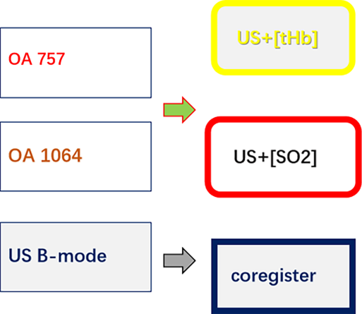
LOUISA-3D system imaging mode
Laser Optoacoustic Ultrasonic Imaging System Assembly for enhanced breast cancer detection and diagnostics


- Full View 3D
- US + OA
- 5 min scan
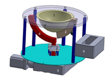
- 1.Breast Cup (disposable)
- 2.Array of Transducers
- 3.Rotational Stage
- 4.Fiberoptic Illuminator
- 5.Step Motor
- 6.Pre-Amplifier
-
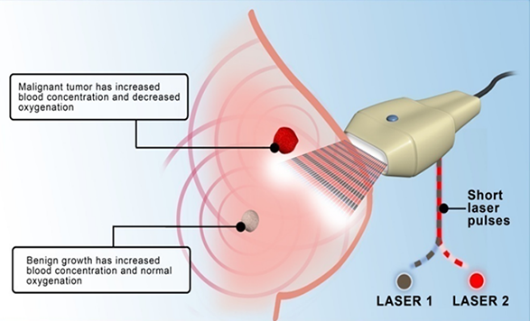
Coregistered Functional+Anatomical OA Imaging for detection and differentiation of breast tumors
Using endogenous contrast agent hemoglobin to achieve imaging of breast blood vessels and breast tumors
Classification of breast cancer, boundary determination of breast tumor, quantitative determination of focal hemoglobin concentration and blood oxygen saturation
It provides a new tool for evaluating the effect of chemotherapeutic drugs on breast cancer treatment and for tracking and diagnosing breast lesions that cannot be identified by conventional imaging techniques
 Oraevsky et al. Photoacoustics 2018; 12: 30-45[THb]=[Hb]+[HbO2][SO2]=[HbO2]/[THb]
 -
Coregistered OA-US Imaging for Guiding Biopsy and Surgery

- While locating biopsy clips on ultrasound is challenging, these biopsy clips can be easily detected with real-time optoacoustic imaging
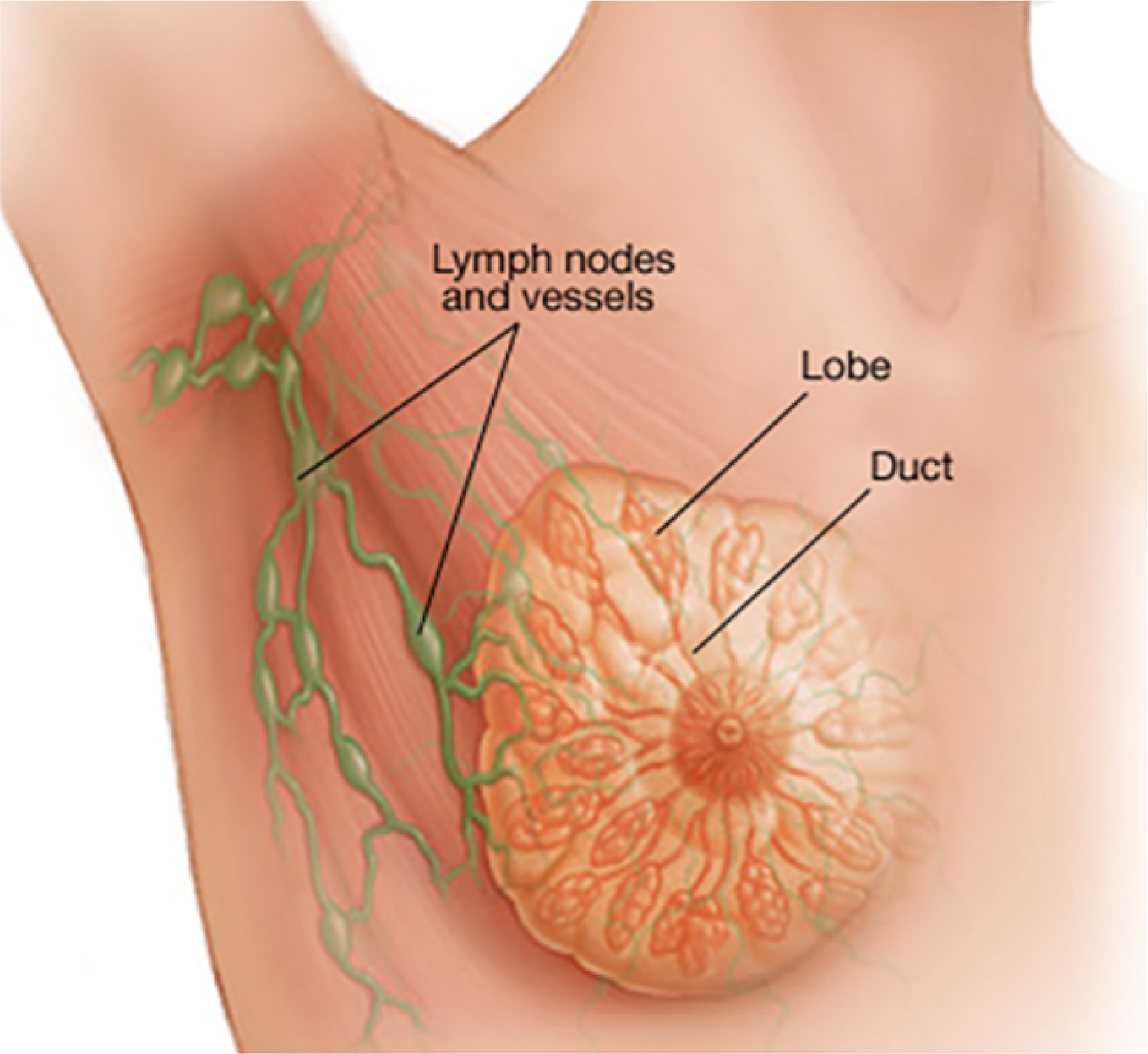
- Lymph nodes can located by ultrasound and targeted with agents for diagnostic imaging of cancerous nodes using optoacoustics
LOUISA-3D Image of [tHb] in a normal patient Breast


 Present position: